


1. Research Theme 2 Development of “Tissue Penetrating Cell Technology” and “in vivo Imaging Methods”
- (1)Tissue penetrating cell analysis, drug targeting using these cells’ properties
and application in disease treatment - (2)Development of innovative bio-imaging methods
- In vivo observation of gene expression using MRI for small animals
- Bio-imaging using wide-spectrum near-infra red light sources
- Bio-imaging of toxic transformation of microglia - (3)Controlling cell activation using light
Tissue penetrating cell analysis, drug targeting using these cells’ properties and application in disease treatment
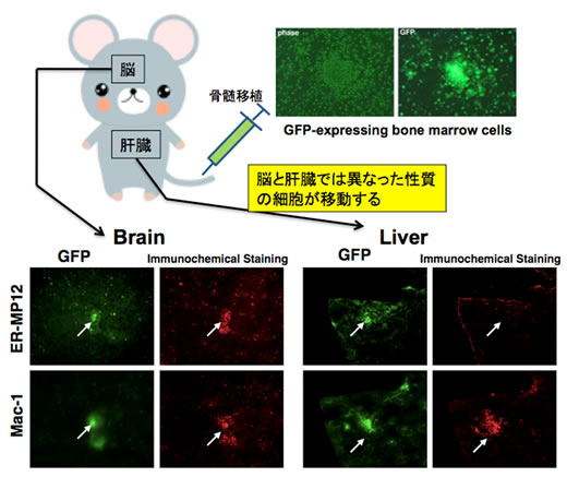
We have found that there are cells in the bone marrow that neuro-selectively migrate in the same way as microglia, even though there are very few of these cells. They retain undifferentiated properties in the normal mouse brain. However, these cells have differentiating and accumulating properties in response to injuries or inflammation in the brain. We are looking into what kind of response these cells have and the roles they play in response to brain diseases. In addition to working on the brain, we have discovered the presence of cells that exhibit selective migration in specific peripheral tissues, and we are looking into the interactions between cells in various diseases by controlling the activation of these cells, with the aim of using this in future therapy.
(2) Development of an innovative bio-imaging method
We are using MRI for small animals, light imaging technology, PET, microscopy imaging, and similar technologies, to develop technology to visualize changes in animals occurring at the organs and cellular levels in a minimally-invasive manner, as well as to carry out research into biological phenomena.
In vivo observation of gene expression with small animal MRI: We focused on the fact that the iron-storage protein ferritin enhances a negative imaging effect on T2 high-contrast MRI images, and, by making it expressed in cells under the control of a specific promoter, we developed a method for visualizing the movement of cells inside organisms based on gene expression.
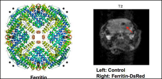
Image on left: The 3-D structure of ferritin. A single ferritin molecule can adsorb 4500 iron atoms.
Image on right: It is possible to visualize the distribution of ferritin proteins using MRI imaging
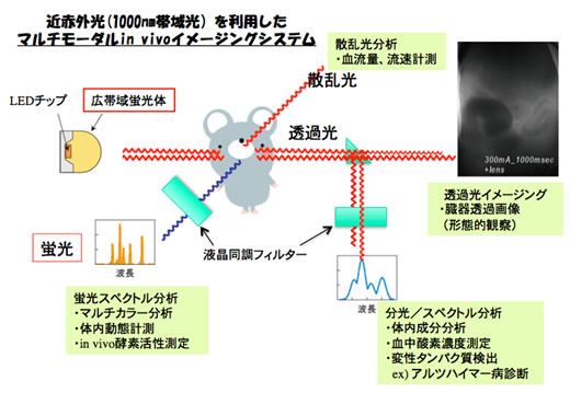
Biological imaging using wide-area near infrared light sources: We have developed a method of imaging biological functions using near infrared light, which passes through biological matter, with a new broad-spectrum fluorescent material, in collaborative research with the Graduate school of Engineering. It is possible to analyze biological components, observe disease states, and analyze biochemical changes due to disease, by using the various properties of light.
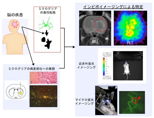
Bio-imaging of toxic transformation of microglia: Microglia get activated when neurons and other cells degenerate due to various diseases. They aggregate at the site of disease, and some of them proliferate. Microglia, which aggregate in an activated condition, either attack and exclude cells with irreversible functional failure (cytotoxic microglia), or they have the role of protecting other cells from damage (protective microglia). Early diagnosis of various diseases such as Alzheimer’s, Parkinson’s, stroke, and brain tumors may be possible if there is a way of identifying the state of these microglia. Activated microglia cause certain characteristic changes, one of which is a change in the strength of ligand binding to peripheral benzodiazepine receptors. It has been suggested that this change may be closely related to the state of activation of microglia, which is closely related to legion sites. We carried joint research with the Faculty of Radiological Technology, School of Medicine at the Fujita Health University, and the Research Institute, National Institute of Geriatrics and Gerontology, the University of Toronto in Canada, and the University of Bari in Italy and have been carrying out research on imaging ligands (PET ligands and fluorescent probes) which can distinguish between protective and cytotoxic microglia. It is considered that if this research makes it possible to distinguish between cytotoxic and protective microglia, it will be not only possible to detect pathological development at an early stage, but also to estimate the severity and prognosis of these diseases.
(3) Control of cell activation using light
Having brain cells express light-sensitive ion channels, we have been carrying out research on adjusting brain and organism functions, by controlling the condition of cells with irradiation by light.
The membrane protein called Channelrhodopsin-2 (Chr2), which is derived from Chlamydomonas algae, is known to be an ion channel which selectively responds to blue light. Since the activity of Chr2 is not mediated by the cell matrix, unlike the Rhodopsin group in mammalian cells, it is possible to directly control the opening and closing of this ion channel through light stimulation. In recent years, a lot of research has been carried out on nerve circuit networks using neurons expressing Chr2. In addition, we have created not only neurons, but also glial cells to which genes have been transferred, and have been developing the technology to artificially adjust brain functions by activating specific cells.
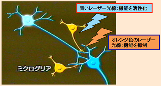
Artificially adjusting the functions of microglial cells and looking into their roles in the brain by switching the genes transferred to the modified microglia on/ off using a laser beam.